Services
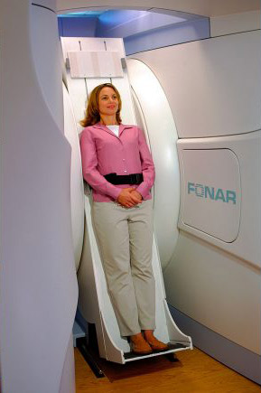
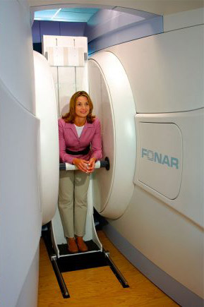
MRI
Last Updated: May 2026
Magnetic resonance imaging (MRI) is a diagnostic imaging system using radio waves, an extremely strong magnetic field, and a computer to visualize internal organs of the human body and obtain diagnostic information. An MRI creates images of the body in thin “slices,” similar to slicing a loaf of bread, allowing doctors to see inside the body layer by layer. Compared to a CT scan, MRI shows differences between soft tissues more clearly.
MRI images are produced without the use of ionizing radiation. The procedure is considered very safe, and significant side effects are rare. The procedure is painless and non-invasive, though you may feel a slight warmth in the area being scanned. You will hear loud tapping or knocking sounds, which is normal as the machine works to capture the images. To ensure your comfort, you will be provided with earplugs or headphones. In some instances, contrast agents, such as gadolinium, are used to enhance certain anatomical structures and increase the diagnostic accuracy of the images.
Magnetic resonance imaging (MRI) is one of the advanced imaging technologies utilized at Akumin and is used to examine virtually all areas of the body. MRI produces highly detailed images of the head, neck, spine, muscles, joints, and bones, and is also commonly used to evaluate the head, spine, joints, abdomen, and pelvis, in addition to veins and arteries of the body.
Akumin offers a range of specialized MRI exams tailored to specific diagnostic needs, including prostate MRI, cardiac MRI, and advanced brain MRI analysis at many of our outpatient imaging centers. Patients living with pain stimulators can also learn about MRI safe scanning options. MRI is especially valuable because it often allows physicians to differentiate abnormal (diseased) tissue from normal tissue more clearly than other imaging modalities such as X-ray, CT, or ultrasound, supporting more accurate diagnosis and personalized care.