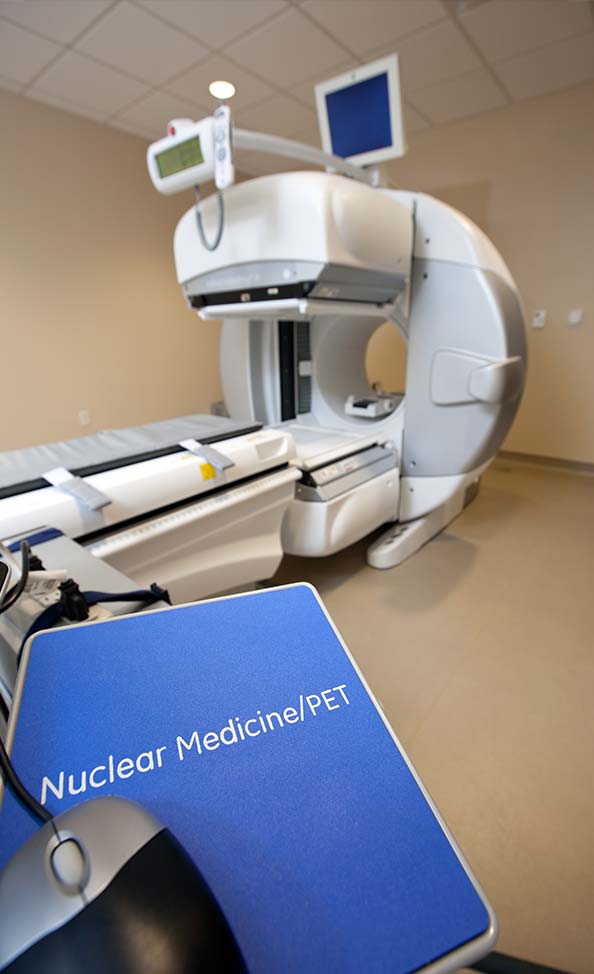
Services

Nuclear Medicine

Last Updated: May 2026

Nuclear medicine is a medical imaging specialty that uses very small amounts of radioactive materials to visualize and measure how organs and tissues function. Rather than focusing only on anatomy, these studies highlight physiology, how the body is working, so clinicians can detect issues earlier, monitor progression, and tailor therapies. At Akumin, nuclear medicine complements radiology exams such as CT, MRI, and ultrasound by adding functional insights that support evaluation of the heart, thyroid, lungs, kidneys, bones, and more.

Nuclear medicine imaging works alongside Akumin’s radiology services—including CT, MRI, and ultrasound to help identify and diagnose many conditions at earlier stages. By showing how organs and tissues perform, nuclear medicine provides important functional detail that complements the anatomic images from other modalities and supports assessment of the heart, thyroid, lungs, kidneys, and bones. As part of our integrated care, these techniques deliver information that guides timely, accurate decision-making.

Nuclear Medicine PET/CT MachineThis specialty uses tiny amounts of radiotracers, which may be injected, inhaled, or swallowed, depending on the study. Our care team selects the appropriate tracer and imaging protocol based on symptoms and medical history, often coordinating nuclear medicine imaging with same-day or follow-up radiology exams to provide a more comprehensive view of a patient’s condition. These coordinated approaches ensure that nuclear imaging findings align seamlessly with a patient’s overall diagnostic plan.

As the tracer travels to specific organs or areas of interest, it emits gamma rays. A specialized camera with advanced software detects this energy to create images that reveal how the body is functioning. These images can be fused with Akumin CT scans in hybrid studies (such as PET/CT) to align functional data with precise anatomy, aiding in the detection and assessment of cancers, heart disease, infections, bone disorders, and thyroid conditions.

Nuclear medicine procedures are generally considered safe and are integral to managing a variety of diseases, using the minimum amount of tracer needed to achieve high-quality imaging. Board-certified technologists and radiologists follow stringent safety and quality standards and collaborate across imaging specialties to guide diagnosis, monitor treatment response, and help plan individualized care.

Condition Application
Kidney dysfunction or obstruction Renal scan to evaluate filtration, drainage, and blood flow
Thyroid disorders or nodules Thyroid scan to measure uptake/function and characterize nodules
Arthritis, bone pain, or suspected tumors Bone scan to detect inflammation, fractures, lesions, or metastases
Infection or inflammation of unknown origin Gallium scan to localize infectious or inflammatory processes and abscesses
Coronary artery disease or evaluation after a heart attack Heart perfusion/function scans to assess blood flow and ventricular performance
Cerebrovascular or neurologic abnormalities Brain scan to evaluate regional blood flow or focal abnormalities
Breast cancer evaluation Breast-specific imaging used with mammography to assess suspicious areas

What to Expect During a Nuclear Medicine Scan

Your technologist will review your medical history and explain the exam. A small amount of radiotracer is administered by IV, inhalation, or swallowing, then time is allowed for the tracer to collect in the target organ or tissue. You’ll lie on an imaging table while a gamma camera (or PET/SPECT system) acquires images, typically over 20 to 60 minutes, depending on the study. You may be asked to remain still, change position, or return for delayed images. After the scan, you can resume normal activities, and drinking fluids often helps clear the tracer from your body. For certain indications, a PET/CT or a SPECT scan may be performed to capture additional functional detail.


Frequently Asked Questions (FAQS)

Is nuclear medicine safe?

Yes. The amount of radiation used is typically comparable to or lower than many diagnostic imaging exams. Your care team tailors the dose to your body and clinical needs and follows strict safety guidelines.

How should I prepare?

Preparation varies by exam. You may be asked to avoid certain foods, caffeine, or medications, or to fast for a short period. Detailed instructions will be provided when your appointment is scheduled, including any preparation specific to nuclear imaging such as for a PET/CT or a SPECT scan.

Will I need someone to drive me home?

Most exams do not require sedation, so you can usually drive yourself and return to normal activities right after the scan unless instructed otherwise.

How long does the visit take?

Plan for 1 to 3 hours, depending on the study and whether delayed images are needed. Imaging time itself is often 20 to 60 minutes.

When will I get results?

A board-certified radiologist interprets your images and sends a report to your referring provider, who will review the results with you. Turnaround is typically within a few business days.

Can I be around family after my scan?

In most cases, normal contact with others is safe. Drinking fluids and frequent urination can help clear the tracer more quickly. Your technologist will share any special precautions if needed.

What if I’m pregnant or breastfeeding?

Please inform the team before your exam. Certain studies may be postponed or adjusted to ensure safety for you and your child.

Will my insurance cover the exam?

Many plans cover medically necessary nuclear medicine studies. Akumin can help verify benefits and discuss any expected out-of-pocket costs prior to your appointment.

What is the difference between MRI and nuclear medicine?

MRI uses strong magnets and radio waves to create detailed pictures of anatomy and soft tissues without ionizing radiation; it shows structure. Nuclear medicine uses radiotracers and specialized cameras to show organ function and metabolism; it highlights physiology. These approaches answer different clinical questions and are often complementary.

What should I avoid after a nuclear medicine scan?

Most patients can resume normal activities. It’s helpful to drink plenty of fluids for the next 24 hours to flush the tracer. Unless you receive specific instructions, you do not need to avoid routine contact; however, as a precaution, you may choose to limit prolonged close contact with infants and pregnant individuals for several hours. Follow any exam-specific guidance provided by your technologist.

Contributors

Bhea Jaime, CNMT
Radiation Safety Specialist

Bhea is a healthcare leader with a strong technical background in Nuclear Medicine, PET/CT, radiopharmaceutical therapies and radiation safety in clinical and hospital settings. She brings experience in regulatory compliance, oversight of radiation safety and treatment programs across multi-site healthcare systems, with a focus on clinical team collaboration and operational excellence.

Request an Appointment

Complete the form below to request an appointment. Kindly note that not every procedure is available at all of our locations. To search for all procedures available in your area visit our locations page.

Request An
Appointment

Online or over the phone, we make it easy to schedule your exam

Book now ›

Find Your
Center

Now we’re closer than ever with 100+ fixed-site centers nationwide

Locate now ›

Contact Us For
Medical Questions

Speak with our clinical team about CT procedures, prep, and more

Contact now ›